
Unlike live cell therapies, Cellmax™ delivers bioactive signaling proteins without the risks of immune rejection or tumorigenicity.

Redirection: Shifts macrophages from destructive M1 pro-inflammatory states to reparative M2 healing states.

Stabilization: Suppresses damaging pro-inflammatory cytokines (e.g., IL-1β, IL-6), resolving hyper-inflammation and restoring structural integrity.
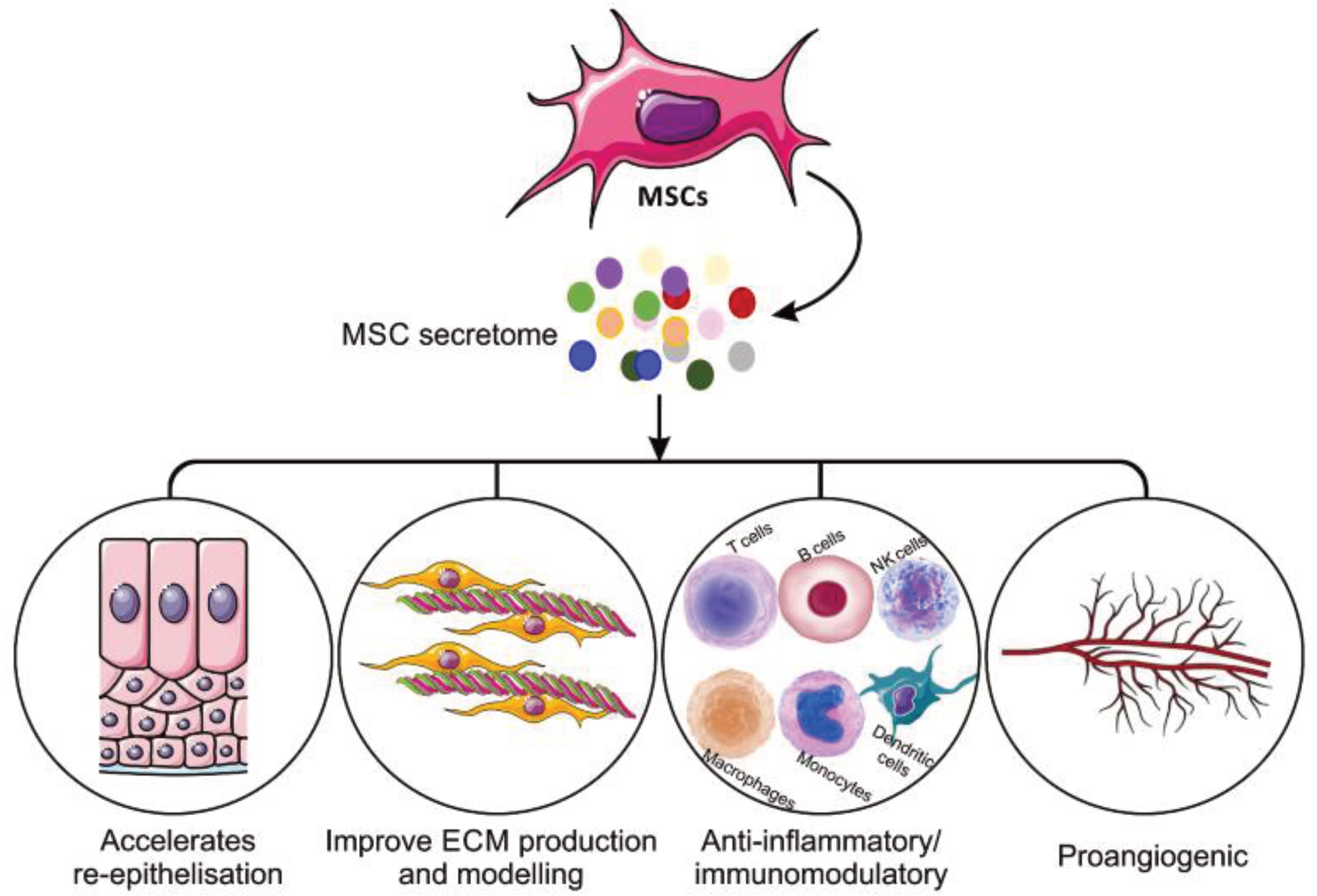
Bioprotein has been identified as one of the main contents of Cellmax™. This bioprotein contains activated growth factors that support skin repair, regenerate cells, induce vascularizations and promote skin rejuvenation.

Bioprotein Content
Function
Chemokines Direct migration of white blood cells to damaged or ruptured tissues
Pro-inflammatory Cytokines Regulate growth, cell activation, differentiation and homing of the
immune cells to the sites of infection
Anti-inflammatory Cytokines Control pro-inflammatory response
Growth Factors
Fibroblast Growth Factor 2 (FGF2) Prevents endothelial cells from undergoing cell death and promotes endothelial cell proliferation and angiogenesis
Transforming Growth Factor Beta 1 (TGFβ1) Stimulating angiogenesis, fibroblast proliferation, collagen synthesis and deposition and remodeling of the new extracellular matrix
Vascular Endothelial Growth Factor A (VEGF A) Stimulates wound healing through angiogenesis and promotes collagen deposition and epithelialization

Learn how to use Cellmax™ Freeze Dried Powder and the application method
(For use by Healthcare Professionals only)

Application Method:

  1. All previous dressings must be removed. Follow standard wound care procedure including wound debridement.
  2. Wound debridement: removal of dead, necrotic material, slough, damaged, or infected tissue. Removal may be surgical based on standard wound care technique.
  3. For patients with low pain tolerance, apply a local anesthetic (e.g., 5% lidocaine gel) for 10–15 minutes and wipe off before administration.
  4. Apply Cellmax™ to the wound bed or peri-wound area, followed by non-absorbable primary and secondary dressings.
  5. Administer weekly for up to 12 weeks or until wound closure, with wound assessments at each visit.

Reconstitution (Freeze Dried Powder):

  1. Inspect vial integrity. Disinfect rubber stopper with sterile alcohol swab for 15 seconds and allow to air dry.
  2. Withdraw 2 mL sterile 0.9% Normal Saline and inject into the vial.
  3. Gently swirl until fully dissolved and clear. Do not shake vigorously.
  4. Use immediately or store at 2–8°C for up to 12 hours (single-patient use; discard unused solution).

Subcutaneous Injection:

  • Inject at 1.0–3.0 mm depth into the subcutaneous layer at a 45° angle from the wound surface.
  • Begin approximately 3 mm from the wound edge.
  • Maintain ~1 cm spacing between injection sites around the periwound.

Wound Spray Application:

  • Shake gently before use. Spray evenly over the wound bed, 1–2 sprays per cm².
  • Apply 2–3 times per week for up to 12 weeks.
  • Store at 2–8°C after first use. Discard within 28 days.
  • Safer and superior alternative to live-cell therapies
  • Eliminates risks like immune rejection and tumorigenicity
  • Precisely resets the wound microenvironment and molecular signals
  • Non-synthetic medical therapy approach to wound healing
  • Expedite wound recovery for patients with comorbidities
  • High potency, stable and easy to store
  • Free of cells
  • Cell-specific therapeutic effects

Indications

Cellmax™ products are indicated as an adjuvant therapy to standard wound care for healthcare professionals in managing acute, chronic and complex wounds, including:

  • Diabetic Foot Ulcers (DFU)
  • Venous Leg Ulcers (VLU)
  • Postoperative Wounds
  • Traumatic Wounds
  • Burn Wounds
  • Bedsores
  • Wounds with compromised peri-wound skin (HSPC Class 2–3)(Applies to all age groups)

Cellmax™ offers a paradigm shift in wound care manegement with the emerging role of MSCs-derived secretome in cell-free therapy.

Cell-free therapy uses secreted products (secretome) from stem cells as an innovative approach to promote several biological functions, such as angiogenesis, neovascularization and arteriogenesis¹.

Secretome is defined as a range of bioprotein and bioactive substances produced by a cell in the extracellular space, which includes but is not limited to proteins, nucleic acids, proteasomes,exosomes, mciroRNA and membrane vesicles².

Cellmax™ provides an excellent treatment option for wound care management by accelerating angiogenesis, fibroblast proliferation, collagen synthesis and epithelization.

References
  1. Koike, Y. et al. (2020). Fibroblast growth factor 2 accelerates the epithelial–mesenchymal transition in keratinocytes during wound healing process. Sci Rep 10, 18545.
  2. Barrientos, S. et al. (2008). Growth factors and cytokines in wound healing. Wound Repair and Regeneration, 16: 585-601.
  3. Bao, P. et al. (2009). The role of vascular endothelial growth factor in wound healing. The Journal of Surgical Research, 153(2), 347–358.
  4. Ferrara, N. et al. (1992). Molecular and biological properties of the vascular endothelial growth factor family of proteins. Endocrine reviews, 13(1), 18–32.
  5. Abbasi-Malati, Z. et al. (2018). Mesenchymal Stem Cells on Horizon: A New Arsenal of Therapeutic Agents. Stem Cell Reviews and Reports, 14.
  6. Eleuteri, S. & Fierabracci, A. (2019). Insights into the Secretome of Mesenchymal Stem Cells and Its Potential Applications. Int J Mol Sci, 20.
  7. Nair, H. K. R. et al. (2025). A Quasi-experimental Study of Cellmax™ as an Adjuvant Therapy for Individuals with Chronic Wounds: Clinical Investigation Report, V2. CIP: CELLMAX-23-1.6; Ethics Ref: NMRR ID-23-00821-PEC; MDA Notification: MDA/CIU/2024/6370.

Image reproduced under an open access license from Ahangar et al., Copyright 2020, © Authors. Ahangar, P.; Mills, S.J.; Cowin, A.J. Mesenchymal stem cell secretome as an emerging cell-free alternative for improving wound repair. Int. J. Mol. Sci. 2020, 21, 7038.